Conjoint Tendon Shoulder Anatomy / Axilla: Anterior View | Shoulder anatomy : It gets its name from the fact that it is often continuous or conjoined with the tendon of the internal oblique, another of the abdominal muscles.
Conjoint Tendon Shoulder Anatomy / Axilla: Anterior View | Shoulder anatomy : It gets its name from the fact that it is often continuous or conjoined with the tendon of the internal oblique, another of the abdominal muscles.. Shoulder muscles and shoulder tendons. Shoulder radiology & anatomy at usuhs.mil. Muscles allow us to move by pulling on bones. Ligaments are soft tissue structures that connect bones to bones. An introduction to the anatomy of the shoulder.
Shoulder tendonitis is the inflammation, irritation and swelling of the tendons in the rotator cuff and bicep. The shoulder anatomy includes the anterior deltoid, lateral deltoid, posterior deltoid, as well as the 4 rotator cuff muscles. Related online courses on physioplus. Robin smithuis and henk jan van der woude. Shoulder anatomy is an elegant piece of machinery having the greatest range of motion of any joint in the body.
Axilla: Anterior View | Shoulder anatomy from i.pinimg.com The shoulder joint (glenohumeral joint) is a ball and socket joint between the scapula and the in this article, we shall look at the anatomy of the shoulder joint and its important clinical correlations. The muscle belly then crosses the entire upper arm and separates into two tendons. Specifically, the four rotator cuff muscles include the following In the shoulder it's commonly more than just one structure that gets affected. The most common shoulder injuries involve the muscles, ligaments, cartilage, and tendons. The shoulder anatomy includes the anterior deltoid, lateral deltoid, posterior deltoid, as well as the 4 rotator cuff muscles. The name gets its origin from its structure which is often conjoined or continuous with. Shoulder muscles and shoulder tendons.
These are the main ligaments that help to stabilize the joints of.
(inguinal aponeurotic falx labeled at lower left.) falx (disambiguation) — other parts of the anatomy with names including falx. These are the main ligaments that help to stabilize the joints of. Related online courses on physioplus. The rotator cuff is a group of four muscles and tendons that surround the glenohumeral joint. Anterior graphic of the shoulder. Specifically, the four rotator cuff muscles include the following The name gets its origin from its structure which is often conjoined or continuous with. It gets its name from the fact that it is often continuous or conjoined with the tendon of the internal oblique, another of the abdominal muscles. One tendon might have it worse, but it's never isolated to just one tendon. The long head of biceps (lhb) is a very important tendon that travels through the shoulder joint (glenohumeral joint). Rotator cuff, a network of muscles and tendons that cover the top of the humerus, or upper arm bone, to hold it place and enable the arm to rotate. First, you need to warm up and stretch the shoulder area before any sport or activity that is going to place strain on. Shoulder tendonitis is the inflammation, irritation and swelling of the tendons in the rotator cuff and bicep.
Shoulder radiology & anatomy at usuhs.mil. The muscle belly then crosses the entire upper arm and separates into two tendons. There are several important ligaments in the shoulder. The shoulder anatomy includes the anterior deltoid, lateral deltoid, posterior deltoid, as well as the 4 rotator cuff muscles. Shoulder muscles and shoulder tendons.
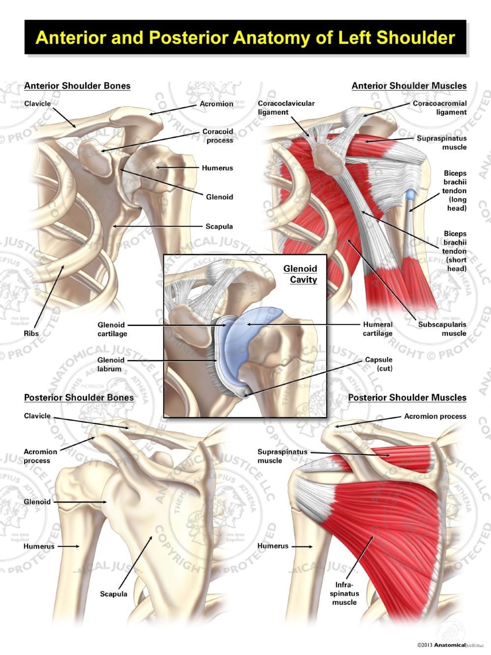
Anterior and Posterior Anatomy of the Left Shoulder ... from anatomicaljustice.com There are several important ligaments in the shoulder. The shoulder is made up of three bones: The scapula (shoulder blade), clavicle (collarbone) and humerus (upper arm bone). Robin smithuis and henk jan van der woude. Normal anatomy, variants and checklist. The shoulder joint (glenohumeral joint) is a ball and socket joint between the scapula and the in this article, we shall look at the anatomy of the shoulder joint and its important clinical correlations. The shoulder is comprised of a ball (humerus) and socket (scapula), bones, ligaments, tendons and muscles that move the arms and connect them to the torso. This article covers the anatomy of the inguinal canal, including contents, borders and related clinical aspects, such as hernias.
Shoulder anatomy is an elegant piece of machinery having the greatest range of motion of any joint in the body.
The muscle belly then crosses the entire upper arm and separates into two tendons. ^ clinical anatomy by ernest w. These are the main ligaments that help to stabilize the joints of. The scapula (shoulder blade), clavicle (collarbone) and humerus (upper arm bone). There are a few ways you can help prevent shoulder tendonitis occuring. The tendons are the attachment of the. Shoulder tendonitis is the inflammation, irritation and swelling of the tendons in the rotator cuff and bicep. Tendons are fibrous cords attached to muscles and bone. Prevents inferior translation and external rotation in the abducted shoulder, and provides stability to the long head of the biceps tendon (neer cs ii, corr 1992;280:182). One tendon might have it worse, but it's never isolated to just one tendon. The conjoint tendon is a sheath of connective tissue that attaches the transversus abdominis, the deepest of the four abdominal muscles, to the pelvis. Your biceps tendons attach the biceps muscle to bones in your shoulder and in your elbow. Shoulder anatomy for ultrasound evaluation.
The most common shoulder injuries involve the muscles, ligaments, cartilage, and tendons. The conjoint tendon (previously known as the inguinal aponeurotic falx) is a structure formed from the lower part of the common aponeurosis of the internal oblique muscle and the transversus abdominis as it inserts into the crest of the pubis and pectineal line immediately behind the superficial inguinal ring. Shoulder anatomy for ultrasound evaluation. There are a few ways you can help prevent shoulder tendonitis occuring. Upper limb trauma programme injuries.
Conjoint Tendon Shoulder Anatomy - Best Y1s2 Gi Block ... from www.uptodate.com It reduces wear and tear. Shoulder tendonitis is the inflammation, irritation and swelling of the tendons in the rotator cuff and bicep. This article covers the anatomy of the inguinal canal, including contents, borders and related clinical aspects, such as hernias. (inguinal aponeurotic falx labeled at lower left.) falx (disambiguation) — other parts of the anatomy with names including falx. The biceps muscle has two tendons at the shoulder, called the long head and short head. Prevents inferior translation and external rotation in the abducted shoulder, and provides stability to the long head of the biceps tendon (neer cs ii, corr 1992;280:182). The shoulder joint is the connection between the chest and the upper extremity. It gets its name from the fact that it is often continuous or conjoined with the tendon of the internal oblique, another of the abdominal muscles.
Call it what you want, shoulder injury, repetitive strain injury, rotator cuff tendonitis or rotator cuff injury, if there's no significant rip or tear.
Ligaments are soft tissue structures that connect bones to bones. The conjoint tendon can be describe as a layer of connective tissue which connects the pelvis to the transversus abdominis, the deepest of the 4 muscles of the abdomen. Muscles allow us to move by pulling on bones. The long head of biceps (lhb) is a very important tendon that travels through the shoulder joint (glenohumeral joint). (inguinal aponeurotic falx labeled at lower left.) falx (disambiguation) — other parts of the anatomy with names including falx. Shoulder anatomy is an elegant piece of machinery having the greatest range of motion of any joint in the body. There are a few ways you can help prevent shoulder tendonitis occuring. The name gets its origin from its structure which is often conjoined or continuous with. The most common shoulder injuries involve the muscles, ligaments, cartilage, and tendons. The shoulder joint is the connection between the chest and the upper extremity. Shoulder anatomy for ultrasound evaluation. Rotator cuff, a network of muscles and tendons that cover the top of the humerus, or upper arm bone, to hold it place and enable the arm to rotate. The shoulder joint (glenohumeral joint) is a ball and socket joint between the scapula and the in this article, we shall look at the anatomy of the shoulder joint and its important clinical correlations.
The conjoint tendon is a sheath of connective tissue that attaches the transversus abdominis, the deepest of the four abdominal muscles, to the pelvis shoulder tendon anatomy. The conjoint tendon then turns inferiorly and attaches on.


0 Comments